

Recent HRIA research, conducted in partnership with medical universities, aims to improve diagnostic accuracy through neural architectures optimized for various types of scans (CT, MRI, X-ray, OCT).
Vascular Network Segmentation and Radiographic Analysis
Structural improvements are introduced to standard models to increase sensitivity in detecting fine anatomical structures:
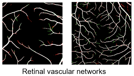
Angio-OCT (Retinal Networks): The I-MedSAM model was used, augmented with a Frangi module. This modification led to a +1% improvement in Dice, Precision, and Sensitivity metrics for vascular network segmentation.
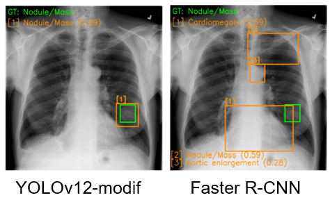
Chest Radiographs: An improved YOLO_V12 architecture was developed for detecting thoracic pathologies (cardiomegaly, aortic dilation, pulmonary nodules, and pleural effusion).
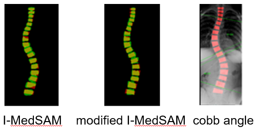
Scoliosis Assessment: Use of YOLO OBB (Oriented Bounding Boxes) combined with a modified I-MedSAM model for automatic Cobb angle calculation.
Tumor Recognition through Domain Adaptation Learning (DAL)
A major contribution consists in the use of domain adaptation learning techniques for liver and pancreatic tumor recognition:
- Dataset: Contrast-enhanced CT images from patients with liver tumors (45 patients) and pancreatic tumors (30 patients).
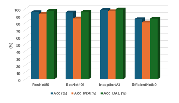
- Methodology: A mixed dataset was used, followed by fine-tuning through DAL.
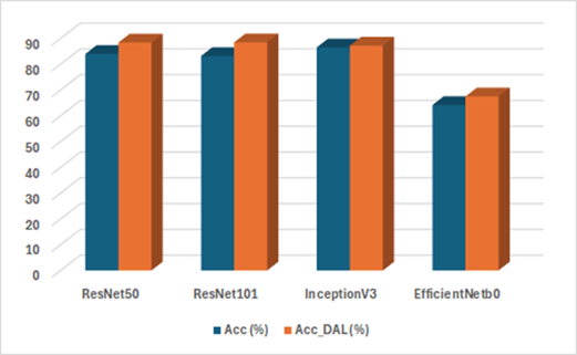
- Performance: Models such as ResNet50, ResNet101, InceptionV3, and EfficientNetB0 were evaluated comparatively. The results indicate that the DAL approach (Acc_DAL) consistently outperforms the accuracy of models trained in a standard way or on mixed datasets.
Research Directions and Assisted Diagnosis (UMF Partnership)
In collaboration with UMF Cluj, AI-assisted diagnostic systems are being developed for complex liver pathologies:
- Focal Lesions: Classification of hepatic lesions using abbreviated MRI protocols.
- Hepatic Fibrosis: Classification of fibrosis stage through multimodal analysis (CT/MRI/Ultrasound).
- Treatment Monitoring: Evaluation of treatment response for hepatic lesions using CT scans.
- Portal Hypertension: Automated diagnosis of portal hypertension using CT data.